| dc.contributor.author | Öztekin, Pelin Seher | |
| dc.contributor.author | Yılmaz, Behice Kaniye | |
| dc.contributor.author | Gökharman, Fatma Dilek | |
| dc.contributor.author | Koşar, Pınar Nergis | |
| dc.date.accessioned | 2019-05-10T09:39:17Z | |
| dc.date.available | 2019-05-10T09:39:17Z | |
| dc.date.issued | 2014 | |
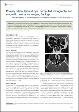
| dc.identifier.citation | Öztekin, P. S., Yilmaz, B. K., Gokharman, F. D., & Koşar, P. N. (2014). Primary orbital hydatid cyst: computed tomography and magnetic resonance imaging findings. Singapore medical journal, 55(11), e184. | en_US |
| dc.identifier.issn | 0037-5675 | |
| dc.identifier.uri | https://doi.org/10.11622/smedj.2014167 | |
| dc.identifier.uri | https://hdl.handle.net/11491/657 | |
| dc.description.abstract | Orbital hydatid cyst is a rare form of hydatidosis, comprising less than 1% of all hydatid cysts reported. The first choice of treatment for orbital hydatid cyst is surgery. Preoperative diagnosis is important, so as to avoid rupture of the cyst and prevent the spread of the parasitic disease. Herein, we present the computed tomography and magnetic resonance imaging findings of a case of primary orbital hydatid cyst. © 2014, Singapore Medical Association. All rights reserved. | en_US |
| dc.language.iso | eng | |
| dc.publisher | Singapore Medical Association | en_US |
| dc.relation.isversionof | 10.11622/smedj.2014167 | en_US |
| dc.rights | info:eu-repo/semantics/openAccess | en_US |
| dc.subject | Echinococcus Granulosus | en_US |
| dc.subject | Hydatid Cyst | en_US |
| dc.subject | Orbita | en_US |
| dc.title | Primary orbital hydatid cyst: Computed tomography and magnetic resonance imaging findings | en_US |
| dc.type | article | en_US |
| dc.relation.journal | Singapore Medical Journal | en_US |
| dc.department | Hitit Üniversitesi, Tıp Fakültesi, Dahili Tıp Bilimleri Bölümü | en_US |
| dc.identifier.volume | 55 | en_US |
| dc.identifier.issue | 11 | en_US |
| dc.identifier.startpage | e184 | en_US |
| dc.identifier.endpage | e186 | en_US |
| dc.relation.publicationcategory | Makale - Uluslararası Hakemli Dergi - Kurum Öğretim Elemanı | en_US |